iDENTical Skull Model with Muscle Attachments M5009
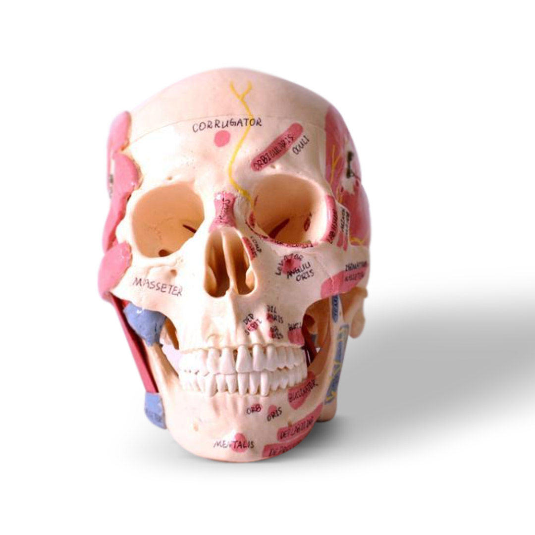
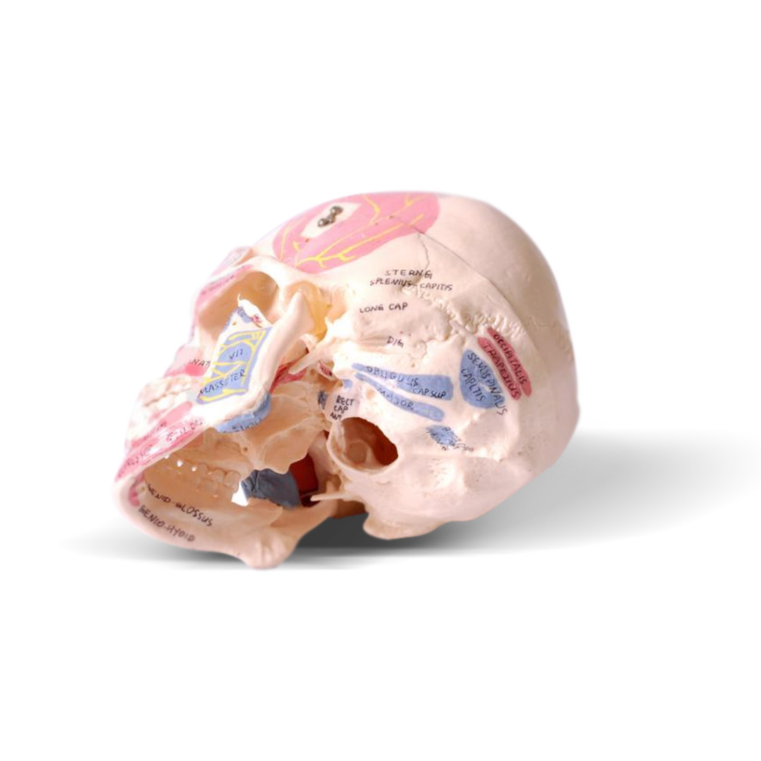
Skull Model Demonstrating various Facial Muscle Attachment
Enter pincode to verify fast clinic delivery & dispatch speed.
Key Highlights
Description
The iDENTical Skull Model with Muscle Attachments is a valuable tool used in medical and dental colleges as well as hospitals for teaching and demonstrating the intricate anatomy of the head and neck region. It provides a comprehensive representation of the muscles involved in mastication, such as the temporalis, masseter, medial pterygoid, and lateral pterygoid muscles. These muscles play crucial roles in jaw movement, including biting, chewing, and grinding food. In addition to the muscles of mastication, the model also displays other important muscles of the head and neck. This includes muscles involved in facial expressions like the frontalis, orbicularis oris, buccinator, zygomaticus major, orbicularis oculi, levator anguli oris, procerus, and mentalis, among many others.
The model’s detailed design allows students to visualize the specific points of attachment of these muscles. The points of origin are highlighted in red, indicating where the muscles originate, while the points of insertion are depicted in blue, representing the areas where the muscles attach. By studying and examining the iDENTical Skull Model with Muscle Attachments, students can gain a better understanding of the complex interactions between muscles and bones in the head and neck region. It helps them grasp the functional anatomy of these structures, aiding in the diagnosis and treatment of various dental and medical conditions.
Key Features
- Accurate Reproduction: The model is a highly accurate reproduction of the human skull, incorporating intricate details and anatomical landmarks. This level of precision allows for a comprehensive understanding of the craniofacial structure
-
Muscle Attachments: The model specifically focuses on muscle attachments, displaying the points of origin and insertion for various muscles. The points of origin are highlighted in red, while the points of insertion are marked in blue, making it easy to identify and differentiate between the two.
-
Visual Aid: The model serves as a visual aid, offering a three-dimensional representation of the skull and muscles. This hands-on approach enhances understanding and retention, enabling students to grasp the spatial relationships between bones and muscles.
-
Durable Construction: The model is constructed using high-quality resin, ensuring durability and longevity. It can withstand frequent handling and use, making it suitable for long-term educational use.
Specifications
- Dimensions: 20cm x 15cm x 13cm
- Material: High-quality resin
- Detailed reproduction of the human skull
- Clear depiction of muscle attachments
- Points of origin highlighted in red
- Points of insertion marked in blue
- Emphasis on muscles of mastication (e.g., temporalis, masseter, medial pterygoid, lateral pterygoid)
- Includes facial muscles involved in expressions (e.g., frontalis, orbicularis oris, buccinator, zygomaticus major, orbicularis oculi, levator anguli oris, procerus, mentalis)
- Three-dimensional visual aid for better understanding
Packaging
- 1 x Skull Model with Muscle Attachments
Directions for Use
- Familiarize yourself with the model: Take some time to examine the model and become acquainted with its features. Identify the different muscles and their corresponding attachments.
-
Study the points of origin and insertion: Pay attention to the red and blue markings on the model. The red points indicate the origins of the muscles, while the blue points represent their insertions. This distinction is crucial for understanding muscle function and movement.
- Explore muscle interactions: Observe how the muscles of mastication, facial expression, and other head and neck muscles interact with the skull. Identify their positions and how they connect to specific bony structures.
-
Manipulate the model: Carefully handle the skull model, rotating it to examine different angles and perspectives. This will allow you to visualize the relationships between the muscles, bones, and surrounding structures.
-
Relate to clinical scenarios: Connect the anatomical knowledge gained from the model to clinical scenarios. Consider how variations in muscle attachments or abnormalities may affect facial expressions, jaw movements, or the diagnosis and treatment of certain conditions.
-
Supplement with additional resources: While the model provides a hands-on learning experience, it can be beneficial to complement your studies with textbooks, anatomy atlases etc. to deepen your understanding of head and neck anatomy.
-
Utilize the model for teaching and demonstrations: If you are an instructor, use the model to teach and demonstrate head and neck anatomy to students. Highlight the important muscle attachments and guide students in their exploration of the model.
Additional Information
Country of Origin: PRC
Manufacturer:
Jining Xingxing Medical Instruments Co., Ltd.
No. 1, Taibai Road, Jining, Shandong, PRC
Packer:
Vasa Denticity Ltd.
Khasra No. 15/1, Mustil No. 6, Village Dabodha, Farrukhnagar, Gurugram, Haryana – 122506, India
Phone: +91 72899 99456
Imported & Marketed By:
Vasa Denticity Ltd.
Khasra No. 15/1, Mustil No. 6, Village Dabodha, Farrukhnagar, Gurugram, Haryana – 122506, India
Phone: +91 72899 99456
Warranty & Service
- Warranty is not applicable for this product.
Package Contents
Standard clinic packaging: 1 x iDENTical Skull Model with Muscle Attachments M5009 (Factory sealed box)
For detailed package breakdowns or bulk master-carton counts, contact our wholesale desk.
| Brand | iDENTical |
|---|---|
| Category | Human Anatomy |
| SKU Code | IDEXX00245 |
| Availability | In Stock (Ready for Dispatch) |
| Product Type | Dental Clinic & Hospital Supply |
Clinical Instructions
- Inspect the packaging seal before clinical application.
- Prepare the operative field following standard clinical isolation protocol.
- Apply according to manufacturer directions and standard restorative technique.
- Dispose of single-use items in bio-medical waste according to healthcare regulations.
Warranty & Return Policy
- 100% Genuine Guarantee: All items are procured directly from verified brand manufacturers & authorized distributors.
- 10-Day Replacement Window: Eligible for return or replacement if received damaged, defective or with broken seal.
- Manufacturer Warranty: Standard warranty applicable according to brand terms.
DentaHaus Benefits
100% Genuine Products
Direct supply from verified dental manufacturers
Best Wholesale Price
Special tiered pricing for clinics & doctors
Fast Pan-India Delivery
Express shipping across all PIN codes
GST Invoice Available
Claim input tax credit on all purchases
Questions & Answers
Get answers from dental experts and verified practitioners
What are the main clinical advantages of iDENTical Skull Model with Muscle Attachments M5009?
Key clinical advantages include ergonomic handling, reliable dimensional stability, compatibility with all standard restorative dental materials, and excellent clinical comfort for patients.
Payment Options
Ratings & Reviews
Verified feedback from practicing dentists & clinics
The material and handling consistency is top-notch. Our clinic orders in bulk batches and we get predictable, durable patient results every single time.
Very convenient product for everyday restorative and clinical procedures. The wholesale 10+ discount saves us substantial clinic supply overhead.
Recommended Products
Still exploring? These dental supplies might fit your clinic needs

iDENTical Typodonts Lower Right Canine For Frasaco Type Jaw Set Individual M8022 – 43
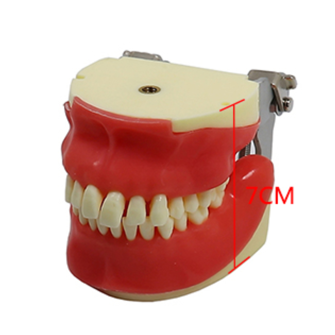
iDENTical Oral Surgery Training Model M4026
iDENTical Oval Resin Block Keychain With Implant Model – Light Green (3023)
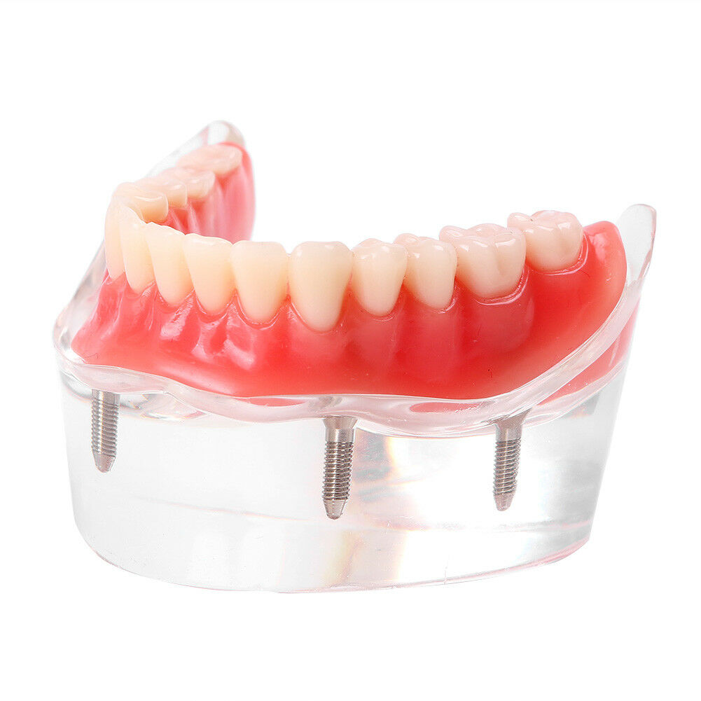
iDENTical Mandibular Overdenture Model with 4 Implants M6003
iDENTical Stethoscope Shape Brooch – Pack of 1 (ZYR-7802)
iDENTical Implant Practicing Maxillary & Mandibular Edentulous Models
iDENTical Third molar Impaction Teeth Model M4009